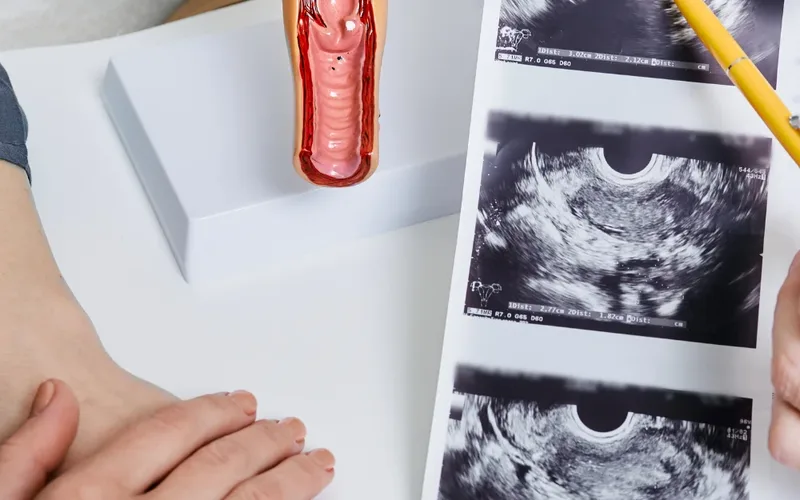
An endometrial biopsy is a short procedure in which a thin, flexible tube is passed through the cervix to take a small sample of the lining of the womb, which is then examined under a microscope. It is most often done to find the cause of abnormal or postmenopausal bleeding, to investigate a thickened lining seen on ultrasound, or occasionally as part of a fertility assessment. Most women feel strong period-type cramping for well under a minute, go home straight afterwards, and have results within one to two weeks.
If your doctor has suggested an endometrial biopsy, you have probably been told it will take only a few minutes, and you are probably more worried about it than that sentence suggests. That is a reasonable reaction. It is an internal procedure, it is usually done while you are awake, and it is being done because something needs to be explained — unusual bleeding, bleeding after the menopause, or a lining that looked thicker than expected on a scan. Being told "it's quick" does not answer the questions that actually matter: why this test, what will I feel, what can the result say, and what happens next.
This page is written to answer those questions in the order you are likely to ask them. An endometrial biopsy is one of the most informative tests in gynaecology precisely because it looks at the cells themselves, and no scan can do that. An ultrasound can tell me how thick your womb lining is and whether it looks even or irregular; only tissue under a microscope can tell me what those cells are doing. Below I explain what the procedure involves, how pain is managed and what can be done if you have found internal examinations difficult before, how to prepare, what each result category means in plain language, and the specific situations in which a sample taken blindly is not enough and we look inside the cavity with a camera instead.
What Is an Endometrial Biopsy?
An endometrial biopsy is the removal of a small sample of the lining of the womb — the endometrium — so that it can be examined under a microscope by a pathologist. It is a diagnostic test, not a treatment. No incision is made and no stitches are needed: the sample is taken through the natural opening of the cervix, which is why it can usually be done in a clinic room in a few minutes rather than in an operating theatre.
The endometrium is the layer that thickens and sheds each month. Oestrogen makes it grow; progesterone, produced after ovulation, matures and stabilises it; when hormone levels fall, it is shed as a period. When that cycle is disrupted — by irregular or absent ovulation, by the hormonal changes of the perimenopause, by weight-related oestrogen production after the menopause, or by certain medicines — the lining can become overgrown or irregular, and the cells themselves can change. The biopsy is how we see those cells.
You may hear the procedure called several different things. Pipelle biopsy or endometrial sampling usually means the office procedure with a thin suction tube. Hysteroscopy-guided biopsy means the sample is taken while a small camera gives a direct view of the cavity. A dilatation and curettage (D and C) is a more complete scraping of the lining, generally done with anaesthesia. All of them answer the same question; they differ in how much of the cavity is sampled and how it is accessed.
It Is Not the Same as a Cervical Smear
This confuses a lot of people, understandably, because both involve a speculum. A smear test samples cells from the surface of the cervix, screening for changes linked to HPV, and the sampler never enters the womb. An endometrial biopsy samples the lining inside the womb, and it is a diagnostic test for a specific problem rather than a routine screening test. If you are trying to make sense of cervical results instead, the meaning of an abnormal Pap smear result is a separate subject with a separate pathway. A normal smear tells you nothing reassuring about the endometrium, and this is a point worth being clear about: women sometimes delay reporting postmenopausal bleeding because their smear was recently normal.
Why an Endometrial Biopsy Is Done
The commonest reason is bleeding that does not fit the expected pattern. The purpose is to rule out or identify precancerous change and cancer of the womb lining, and, when those are excluded, to explain the bleeding so it can be treated properly. The specific situations below each have their own logic.
Bleeding After the Menopause
Any vaginal bleeding after your periods have stopped for a year or more needs to be investigated, however light, however brief, even if it happened only once and even if you are certain it was "nothing". Most postmenopausal bleeding turns out to have a benign cause — most often a thin, fragile, atrophic lining — but a minority, on the order of one in ten, is caused by endometrial cancer. Endometrial cancer is among the most frequently diagnosed gynaecological cancers in many countries, and abnormal bleeding is usually its earliest sign. That combination is exactly why the threshold for investigating is deliberately low.
Heavy, Irregular or Intermenstrual Bleeding Before the Menopause
In women who are still having periods, biopsy is not automatic. It becomes appropriate when bleeding is persistently heavy or irregular, when there is bleeding between periods or after sex, when bleeding has not responded to treatment, or when there are additional risk factors for endometrial change — long-standing cycles without ovulation, polycystic ovary syndrome, a raised body mass index, diabetes, a family history suggesting Lynch syndrome, or treatment with tamoxifen. Age matters too: the same symptom in a woman in her late forties is weighted differently from the same symptom at twenty-five. Many of the hormonal and structural explanations are covered in more detail in the discussion of why periods become irregular.
A Thickened Lining on Ultrasound
Transvaginal ultrasound is almost always the first test. In a postmenopausal woman with bleeding, a lining of 4 mm or less is reassuring and may make biopsy unnecessary, whereas a thicker or irregular-looking lining usually prompts sampling. In women who still menstruate, the lining normally varies a great deal across the cycle, so there is no single number that means "too thick" — which is why measurements are interpreted alongside symptoms rather than on their own. What the numbers mean in each situation is set out in the guide to normal endometrial thickness. One important caveat: if bleeding continues or recurs, a thin lining on an earlier scan does not close the question, and sampling may still be needed.
Unscheduled Bleeding on Hormone Therapy or Tamoxifen
Hormone replacement therapy changes what counts as expected, so it is worth knowing which pattern applies to you. On a sequential, cyclical regimen a monthly withdrawal bleed is planned, and it is bleeding that is unusually heavy, prolonged or at the wrong point in the cycle that needs looking at. On continuous combined therapy, light irregular bleeding in the first three to six months is common while the lining settles, but bleeding that begins later, continues beyond that window, or returns after a bleed-free spell should be assessed rather than attributed to the treatment. Tamoxifen is a different situation again: it acts on the womb lining in a way that thickens and distorts its appearance, so ultrasound measurements are much harder to interpret, and bleeding in a woman taking it is generally investigated with sampling or direct inspection rather than observed.
Before or During Fertility Treatment
Endometrial biopsy is not a routine part of a fertility work-up. It is used selectively — for example where long-standing absence of ovulation has exposed the lining to oestrogen without progesterone for years, where the lining looks abnormal or persistently thickened on scan, or where chronic inflammation of the lining (chronic endometritis) is suspected after repeated implantation failure or recurrent miscarriage. Tests that claim to time embryo transfer by analysing endometrial gene expression are not currently recommended as standard care, and your clinician should be able to explain why a particular test is being offered to you specifically and what will change depending on the answer. The usual sequence of investigations is described under assessment before infertility treatment.
How the Procedure Is Done: Pipelle and Hysteroscopy-Guided Sampling
In the great majority of cases, the sample is taken in an outpatient clinic with a pipelle: a flexible plastic tube about 3 mm across, roughly the width of a drinking straw, passed through the cervix into the cavity of the womb. The appointment lasts ten to fifteen minutes. The part in which the sample is actually taken usually lasts less than a minute.
The Office Pipelle Biopsy, Step by Step
- A pregnancy test is done first if there is any possibility you could be pregnant, because the procedure must not be performed in pregnancy.
- You lie on the couch with your knees bent and apart, covered with a sheet. A speculum is inserted so the cervix can be seen, exactly as for a smear.
- The cervix is cleaned with antiseptic. Local anaesthetic may be applied to or injected around the cervix at this point.
- The pipelle is passed gently through the cervical canal until it reaches the top of the cavity. This is the moment most women feel as a strong cramp.
- The inner piston is drawn back to create suction, and the tube is rotated and moved up and down a few times so that tissue is drawn in from several areas.
- The tube is withdrawn, the speculum removed, and the sample is placed in a preservative solution and labelled for the laboratory.
Occasionally the cervical canal is too tight or too angled for the tube to pass — more common if you have never had a vaginal birth, after cervical surgery, or after the menopause. A thin dilator, a grasping instrument to steady the cervix, medication to soften the cervix beforehand, or ultrasound guidance can solve this. If it still cannot be done comfortably, the sensible answer is not to persist but to arrange the procedure with sedation or under anaesthesia.
Hysteroscopy-Guided Biopsy
Here a hysteroscope — a telescope two to four millimetres wide carrying a camera and light — is passed through the cervix, and a small amount of fluid is used to open the cavity so the whole lining can be seen on a screen. The biopsy is then taken from the exact spot that looks abnormal, and a polyp or small fibroid can often be removed in the same sitting. This can be done awake in an outpatient setting, with or without local anaesthetic, or under sedation or general anaesthesia if the procedure is likely to be longer or if you would prefer to be asleep. Direct vision is the main advantage, for the reasons set out below.
Does an Endometrial Biopsy Hurt, and How Is the Pain Managed?
Most women describe a strong, period-like cramp that builds as the tube enters the womb, peaks for a few seconds while the sample is taken, and fades within a minute or two. Being honest about the range matters more than reassurance: some women find it no worse than a smear, many find it genuinely uncomfortable but manageable, and a minority find it severely painful. Mild cramping often continues for a few hours afterwards and settles with simple painkillers.
Pain is more likely to be significant if you have never given birth vaginally, if your cervix is tight or has been operated on, if you have endometriosis or adenomyosis, if the lining is thin and atrophic after the menopause, if you are very anxious, or if a previous internal examination was painful. None of these are reasons to avoid the test; they are reasons to plan it properly.
What Genuinely Helps
- An anti-inflammatory painkiller taken about half an hour to an hour beforehand, if you can take this class of medicine. Ask which one and what dose is suitable for you.
- Local anaesthetic, applied as gel or spray to the cervix or injected around it as a paracervical block. Availability varies, but you can ask for it, particularly if a previous attempt was painful.
- A slow, narrated technique. Knowing what is happening and when, and being told when the cramping part is about to start, measurably changes how the experience is tolerated.
- Explicit permission to pause or stop. Consent continues throughout the procedure, not just when you sign the form. Saying "stop" is always allowed, and an incomplete attempt can be rescheduled with better pain relief.
- Sedation or general anaesthesia as a planned alternative — appropriate if awake sampling has failed before, if it was intolerable, or if examinations are impossible for you.
If vaginal examinations cause involuntary tightening of the pelvic floor muscles, or are impossible because of pain, say so before the appointment rather than on the couch. That pattern has a name and treatments of its own, discussed under vaginismus and what helps, and the procedure can be arranged around it rather than attempted and abandoned.
One other reaction is worth knowing about because it frightens people unnecessarily: a vasovagal response — feeling suddenly cold, sweaty, nauseated, light-headed or faint while the cervix is being handled. It is a reflex, it is not dangerous, and it passes within minutes once you lie flat with your legs raised. Tell whoever is with you as soon as you feel it coming on.
How to Prepare for an Endometrial Biopsy
Very little preparation is needed: you can eat and drink normally, and you can drive yourself home unless you are having sedation or a general anaesthetic. The important preparation is information, in both directions.
Tell Your Doctor Before the Appointment
- Whether there is any chance you are pregnant, or whether you are trying to conceive and may have conceived this cycle.
- Any blood-thinning medication, including aspirin, or a known bleeding disorder.
- Any current pelvic infection, unusual discharge or pelvic pain.
- All hormonal medication: contraception, hormone replacement therapy, progestogens, tamoxifen or aromatase inhibitors, and when you started them. These change how the lining looks and how the pathologist interprets it.
- Any previous difficult, painful or failed attempt at an internal procedure.
- Whether you have an intrauterine device in place — this needs to be discussed, since it may be left, removed or replaced depending on the circumstances.
When the Procedure Is Postponed
There are only a few reasons to delay rather than proceed. Pregnancy is an absolute one, which is why a test is done whenever there is any doubt. An active pelvic infection is treated first, because passing an instrument through the cervix can carry infection upwards into the womb. A significant bleeding disorder, or full anticoagulation, usually means planning the timing with whoever manages that treatment rather than cancelling the test. None of these change the need for the biopsy; they change only the order in which things are done.
Practical Points
Bring sanitary pads, because light bleeding afterwards is expected. Wear something you will be comfortable in. Timing within the cycle is usually not critical when the question is whether abnormal cells are present, but if the biopsy is being done to assess the hormonal response of the lining, you may be given a specific day to attend. If you are mid-period and bleeding heavily, it is reasonable to ask whether to come on a different day — though in the presence of continuous bleeding the test is done regardless.
Having someone to travel home with is not required for a pipelle biopsy, but many women prefer it, and after sedation it is essential. Most people go back to work the same day. If your job is physically demanding or you know cramping affects you badly, consider arranging the appointment for later in the day. Finally, write your questions down beforehand. Afterwards, while you are crampy and keen to leave, is not when anyone remembers what they wanted to ask.
What the Results Can Show
Endometrial biopsy results fall into a small number of recognised categories, from entirely normal, through benign and treatable conditions, to precancerous change and cancer. The report will use technical words; the explanations below are what they mean in practice. Your result must always be read together with your age, your symptoms, your scan and the medication you take — the same microscopic finding can mean quite different things in a 35-year-old and in a 65-year-old.
Normal and Benign Findings
Proliferative or secretory endometrium describes a lining behaving normally for the first or second half of the cycle. Atrophic or inactive endometrium is the expected thin, quiet lining after the menopause; it is reassuring, and it is itself a common explanation for light postmenopausal bleeding, because a thin lining is fragile. Disordered proliferative endometrium sits in between — an irregular pattern typically seen when ovulation has been infrequent — and is not precancerous, though it often needs treatment for the bleeding. Benign endometrial polyp means a focal overgrowth has been sampled; polyps are a frequent cause of irregular and postmenopausal bleeding and are discussed further under uterine polyps and how they are managed. Chronic endometritis describes persistent low-grade inflammation of the lining, sometimes relevant in recurrent implantation failure, and treated with antibiotics.
Insufficient or Scant Sample
This is common and not a failure on anyone's part. When the lining is very thin, there may simply be almost nothing to collect. In a postmenopausal woman whose scan shows a thin, regular lining and whose bleeding has stopped, an insufficient sample can be accepted as consistent with atrophy. If the lining was thickened or irregular, or if bleeding persists, an insufficient sample is not an answer, and the next step is usually hysteroscopy, as set out in the section on when a biopsy is not enough.
Hyperplasia Without Atypia
The lining is overgrown and crowded, but the individual cells still look normal. This is the consequence of prolonged oestrogen stimulation without the balancing effect of progesterone. It is not cancer. Over years, a small proportion of cases progress, so it is treated — usually with progestogen, often delivered by a progestogen-releasing intrauterine device, alongside attention to reversible contributors such as weight — and a repeat biopsy is arranged, typically after around six months, to confirm the lining has returned to normal.
Atypical Hyperplasia (Endometrial Intraepithelial Neoplasia)
Here the cells themselves are abnormal. This is a precancerous diagnosis, and it is taken seriously for two reasons: it can progress to cancer, and in a meaningful proportion of cases cancer is already present elsewhere in the cavity but was not captured by the sample. Standard management is referral to a gynaecological cancer service, and removal of the womb is usually recommended once childbearing is complete. For a woman who wants to preserve fertility, treatment with progestogen and close surveillance with repeated biopsies is an established alternative that must be chosen with full understanding of what it involves.
Endometrial Cancer
If cancer cells are found, the report will describe the type and grade. Staging investigations, usually imaging, follow, and treatment is planned by a multidisciplinary team. The reason this test exists, and the reason bleeding after the menopause is investigated promptly, is that endometrial cancer most often announces itself early with bleeding, at a stage when treatment options are at their broadest.
How Long Results Take and What Happens Next
Most endometrial biopsy results are available within one to two weeks. The sample has to be fixed, processed into wax, cut into slices a few microns thick, stained, and then read by a pathologist — a sequence of steps that cannot be hurried without compromising it. Delays beyond two weeks usually have a specific cause: additional special stains, molecular or mismatch-repair testing when cancer is found, or a second opinion from another pathologist on a difficult case. Those are all signs of care, not of bad news, although waiting does not feel that way.
Make Sure You Know How You Will Be Told
Before you leave the clinic, establish three things: when the result is expected, who will contact you, and what number to call if you have not heard by then. Then act on that. Never assume that silence means the result was normal — samples get mislaid, letters go astray, and contact details are mistyped. Chasing a result is not being a nuisance.
If the Result Is Normal
- Normal or atrophic lining with bleeding that has settled: no further tests, but report any new bleeding promptly.
- Normal lining but continuing bleeding: the pipelle may have missed something focal, and hysteroscopy is the usual next step.
Each of the other results described above — a polyp, chronic endometritis, hyperplasia with or without atypia, or cancer — carries its own next step, set out in the previous section. Whatever the result, ask for it in writing and ask what it means for you rather than in general. Two questions are usually worth more than a long explanation: does this result fully explain my symptoms, and what would make you want to look again?
Risks, Aftercare and Recovery
Endometrial biopsy is a low-risk procedure, and the common after-effects are cramping and light bleeding that settle within a few days. Serious complications are uncommon, but you should know what they look like.
What Is Expected
Cramping for a few hours, easing with a simple painkiller or a heat pack. Light bleeding or brown spotting for two to five days, sometimes up to a week or until your next period. Feeling a little shaken for an hour or two, particularly if you had a vasovagal reaction. Most women resume ordinary activity, including work and exercise, the same day or the next.
Uncommon and Rare Problems
Pelvic infection occurs in a small minority and shows itself as increasing pain, fever, or offensive discharge in the days afterwards; it responds to antibiotics but needs to be reported rather than waited out. Perforation of the womb — the instrument passing through the wall — is rare, more likely after the menopause or when the cervix is tight, and usually settles with observation. Heavy bleeding is unusual. A failed procedure, where no sample can be obtained, is not a complication so much as a reason to move to hysteroscopy.
Aftercare
- Use sanitary pads rather than tampons or menstrual cups until the bleeding has stopped — usually two to five days.
- Avoid vaginal intercourse, swimming pools and baths while you are still bleeding; showers are fine.
- Take simple analgesia if you need it, and expect it to be enough.
- Do not douche.
- Remember that the biopsy is not contraception. If you are not using any and you have unprotected sex, you can conceive in that same cycle.
If you are being investigated for bleeding while trying to conceive, ask specifically how the result affects your next steps, because treatment of the lining and fertility treatment sometimes need to be sequenced in a particular order.
When a Biopsy Is Not Enough and Hysteroscopy Is Needed
A pipelle biopsy samples blindly and collects tissue from only part of the lining, so it is excellent at detecting changes that affect the whole cavity and much less reliable for problems confined to one small area. That single fact explains almost every situation in which a hysteroscopy is arranged after a biopsy has already been done.
Hysteroscopy is usually recommended when:
- The sample was insufficient and the lining was thickened or irregular on scan, or bleeding is continuing.
- The biopsy was benign but the bleeding has not stopped. A benign result does not override a persistent symptom.
- The scan shows a focal lesion — a suspected polyp, a fibroid bulging into the cavity, a localised thickening or fluid within the cavity.
- The cervix could not be negotiated and sampling failed, which hysteroscopy under anaesthesia can overcome.
- Atypical hyperplasia is found and the extent of the abnormality needs to be defined before deciding on treatment.
- You are taking tamoxifen and have bleeding, since the appearance of the lining on ultrasound is harder to interpret in this situation.
- Bleeding has recurred months or years after a reassuring biopsy. A previous normal result does not cover a new episode.
The advantage is not only diagnostic. Because the operator can see the cavity, a polyp or a small submucous fibroid can often be removed during the same procedure, which both yields a full specimen for the pathologist and treats the cause of the bleeding. Outpatient hysteroscopy feels broadly similar to a pipelle biopsy, with cramping while the cavity is opened with fluid; where a longer procedure is anticipated, or where you would simply rather be asleep, it is done under sedation or general anaesthesia. If you have had one painful awake attempt, say so — that is useful clinical information, not a complaint, and it should change the plan for the next one.
When to Seek Medical Help
Two separate sets of warning signs matter here: the symptoms that mean you should be assessed in the first place, and the symptoms that mean you should seek help after the procedure.
Symptoms That Should Always Be Assessed
- Any vaginal bleeding or blood-stained discharge after the menopause, however light or brief, even on a single occasion.
- Bleeding between periods, or bleeding after sex.
- Periods that have become much heavier, longer or more unpredictable than they used to be, particularly after the age of forty.
- New bleeding on hormone replacement therapy, or a change from your established pattern.
- Persistent watery, blood-tinged or offensive discharge.
- Heavy bleeding with breathlessness, palpitations or dizziness — have this seen urgently.
After the Biopsy, Contact a Doctor If You Have
- Bleeding heavy enough to soak a pad every hour, or passing large clots.
- A temperature above 38 degrees, or shivering and feeling unwell.
- Offensive-smelling vaginal discharge.
- Pain that is getting worse rather than better after the first day, or pain not controlled by simple painkillers.
- Fainting, severe dizziness, or shoulder-tip pain with abdominal pain — seek urgent assessment.
One last point, and it is the most important one on this page: a normal result is reassuring about the day it was taken, not forever. If bleeding returns after a reassuring biopsy, that is a new symptom and it deserves a new assessment. Conditions of the womb lining are highly treatable when they are found early, and bleeding is almost always the earliest signal. Reporting it promptly is the single most useful thing you can do.
Questions Worth Asking at Your Appointment
Patients who ask a few specific questions tend to leave with a clearer plan — and a biopsy is much easier to tolerate when you understand exactly why it is being done and what the result will change. These are the questions I find most useful.
- What exactly are we trying to rule out in my case, and why now?
- Will the sample be taken blindly, or with a camera giving a direct view? Why that choice for me?
- What pain relief is available, and may I have local anaesthetic?
- What happens if the sample turns out to be insufficient?
- If the result is normal but my bleeding continues, what is the next step?
- Does anything about my medication, my weight or my family history change how this result should be interpreted?
And one practical request that is entirely reasonable to make: ask to be told, during the procedure, when the cramping part is about to begin and when it is finished. Knowing where you are in a short, uncomfortable process makes a genuine difference.
Nothing on this page replaces an individual assessment. Your own history, examination findings and scan determine whether a biopsy is needed, how it should be done and what the result means for you, and those are decisions to make with your own doctor.
References
The following current and independent health sources were used in preparing this article.
- NHS. Hysteroscopy
- NICE. Suspected cancer: recognition and referral (NG12)
- MedlinePlus. Uterine Cancer
- RCOG. Patient information